
Skin Cancer Excision in UK: How Is a Skin Lesion Assessed Before Excision Is Considered?
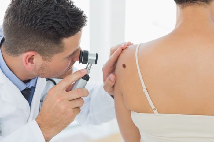
Skin lesions can develop in different forms, sizes, colours and textures. While many skin changes are harmless, some may require further medical assessment to determine whether they could be associated with skin cancer. Understanding the nature of a lesion is an important first step in deciding whether removal, monitoring or further investigation may be appropriate. A careful assessment helps clinicians consider the appearance of the lesion, its development over time and the patient’s medical history before recommending a suitable course of action.
Skin Cancer Excision In UK is a relevant consideration when a lesion has features that require investigation or when a clinician determines that surgical removal may be appropriate. Excision involves removing a lesion, usually with a margin of surrounding skin when indicated, so that the tissue can be examined by a laboratory. However, excision is not automatically recommended for every unusual skin mark. The decision depends on clinical findings, the suspected diagnosis and the most appropriate method of investigation or treatment.
At Well Life Clinic, understanding how a skin concern is assessed can help patients prepare for a consultation and recognise why professional evaluation is important. This article explains the main factors clinicians consider before skin cancer excision is discussed, including changes in appearance, medical history, lesion characteristics and the role of further investigations.
Why Is a Skin Lesion Assessed Before Excision?
A skin lesion is a general term for an area of skin that looks or feels different from the surrounding tissue. It may appear as a mole, patch, lump, scaly area, sore or discoloured mark. Some lesions are present from childhood, while others develop later in life.
The purpose of an assessment is to establish what the lesion might represent and whether it needs further investigation. Clinicians consider whether the change appears consistent with a benign skin condition or whether it has characteristics that could indicate a more serious problem.
Not every suspicious lesion needs the same procedure. Depending on the findings, a clinician may recommend a specialist assessment, a skin biopsy, complete excision or another appropriate diagnostic approach. In some circumstances, a lesion may be monitored, although a potentially suspicious lesion should not be left without suitable medical evaluation.
An assessment also helps determine whether the proposed procedure is appropriate for the lesion’s location, size and suspected type. This supports a more informed decision about the next steps.
How Is the Appearance of a Skin Lesion Evaluated?
The visual appearance of a lesion provides important information during an initial examination. A clinician may assess its colour, shape, borders, surface and relationship to the surrounding skin.
Colour and Pigmentation
Skin lesions can be brown, black, pink, red, white or similar in colour to the surrounding skin. Some lesions contain several shades, while others appear relatively uniform.
A change in pigmentation may be relevant, particularly if a mole develops new colours or becomes noticeably darker or lighter. However, colour alone cannot confirm whether a lesion is cancerous. The clinician considers it alongside other visual features and the patient’s history.
Shape and Border
The outline of a lesion can also influence the assessment. Some benign moles have a regular, symmetrical shape and clearly defined borders. Other lesions may have an irregular outline, uneven edges or an asymmetrical appearance.
These characteristics can help identify lesions that require closer examination. Nevertheless, a regular-looking lesion is not necessarily harmless, and an irregular lesion is not automatically cancerous. A clinical assessment is needed to interpret the findings appropriately.
Surface and Texture
The surface of a lesion may be smooth, rough, scaly, crusted, raised or ulcerated. A persistent sore that does not heal, a lesion that repeatedly bleeds or an area that develops a growing crust may require medical evaluation.
The clinician may also examine whether the lesion feels firm, has a raised edge or appears to extend beneath the visible skin surface. These observations help build a broader picture of the concern.
Why Are Changes Over Time Important?
A lesion’s development can be as significant as its current appearance. A mark that has remained unchanged for many years may be assessed differently from one that has recently started growing, changing colour or developing a different surface.
During the consultation, patients may be asked when they first noticed the lesion and whether it has changed in size, shape or appearance. Other relevant details include the development of itching, tenderness, bleeding or a persistent sore.
Photographs taken at different times may help demonstrate changes, especially when the lesion has evolved gradually. However, photographs cannot replace an appropriate clinical examination.
A useful approach is to seek medical advice when a new or existing skin lesion changes noticeably. Early assessment allows the clinician to determine whether further investigation is required rather than relying on appearance alone.
How Does Medical History Influence the Assessment?
A patient’s medical history helps place the skin findings into context. Even when two lesions look similar, the most appropriate next step may differ according to the individual’s background and other relevant factors.
The clinician may ask about previous skin cancers, earlier biopsies, previous removal procedures and any family history of melanoma or other skin cancers. Information about sun exposure, sunburn history and the use of tanning equipment may also be relevant.
Certain medical conditions or treatments that affect the immune system can influence skin cancer risk. Patients should therefore provide an accurate account of their medical history and current medicines.
Previous skin procedures are also worth discussing. For example, a lesion that has been removed before but has returned may require a different assessment from a lesion that has never been investigated. Existing pathology reports, if available, can help the clinician understand earlier findings.
This information does not establish a diagnosis by itself. Instead, it helps determine how the lesion should be examined and whether specialist input is appropriate.
What Role Does Dermoscopy Play in Skin Lesion Assessment?
Dermoscopy is a non-invasive examination technique that uses a specialised magnifying instrument to view structures within the skin that may not be clearly visible to the naked eye.
When appropriate, a clinician may use dermoscopy to examine the pigmentation pattern, border, internal structures and other features of a lesion. This can provide additional information when assessing certain moles and other pigmented skin changes.
Dermoscopy can support clinical decision-making, but it does not provide a definitive diagnosis in every case. Some lesions require a tissue sample for further examination, particularly when clinical findings raise concern.
The need for dermoscopy depends on the lesion and the clinician’s assessment. Where a lesion appears suspicious, further investigation should not be delayed simply because a preliminary examination has been performed.
When Is a Biopsy or Skin Cancer Excision Considered?
Once the lesion has been assessed, the clinician can consider whether tissue sampling or removal is necessary. The decision depends on the suspected condition, the lesion’s location and size, and the most appropriate diagnostic pathway.
A biopsy involves taking a sample of tissue for examination. In some situations, a complete excision may be recommended so that the entire lesion can be assessed. The choice between these approaches depends on clinical considerations and the type of lesion suspected.
Skin Cancer Excision In UK may be considered when removing the lesion is appropriate for diagnosis or treatment. The procedure involves removing the targeted area of skin. Depending on the suspected diagnosis, the clinician may also remove a margin of surrounding tissue.
The removed tissue is generally sent for histopathological examination, in which a specialist examines it under a microscope. The report can help establish the diagnosis and determine whether the lesion has been completely removed.
If skin cancer is confirmed, the next steps depend on the type of cancer, its characteristics and the findings from the pathology report. Some patients may require further excision, a specialist skin cancer service or additional treatment. The appropriate pathway should follow the clinical findings and relevant UK guidance.
How Do Lesion Size and Location Affect the Decision?
The size and location of a lesion can influence the method used to investigate or remove it. A small lesion in an area with sufficient surrounding skin may be managed differently from a larger lesion or one situated on the nose, eyelid, ear or another anatomically sensitive area.
The clinician may consider the amount of tissue that needs to be removed, the direction of the incision and how the wound could be closed. The availability of suitable surrounding skin and the need to preserve function in the affected area may also be relevant.
In some cases, a straightforward excision may be suitable. In others, specialist planning may be required, particularly if the suspected lesion needs a specific surgical approach or if reconstruction may be necessary after removal.
The aim is to select an appropriate procedure based on the clinical requirements rather than the lesion’s size alone.
What Happens During a Consultation at Well Life Clinic?
A consultation provides an opportunity to discuss the skin concern and understand the recommended next steps. The exact process depends on the service required and the findings of the initial assessment.
The clinician may begin by asking about the lesion’s history, including when it appeared and whether it has changed. They may then examine the affected area and review relevant medical information.
If further investigation is required, the clinician can explain the reason for it and whether referral to a dermatologist or specialist skin cancer service is appropriate. Where excision is being considered, the discussion may cover the proposed method, the expected procedure, tissue examination and follow-up arrangements.
Patients can help make the consultation more informative by bringing previous pathology reports, relevant medical records and photographs showing changes over time. They should also mention any previous treatment involving the same area.
If a lesion is suspicious for skin cancer, it is important to establish the appropriate diagnostic and referral pathway. The suitability of a particular clinic or procedure depends on the patient’s clinical needs and the services available.
Can Scar Revision Surgery Be Considered After Skin Excision?
Scar formation is a normal part of the skin’s healing process after an incision or surgical removal. The appearance of a scar can vary according to the location of the wound, the extent of the procedure, individual healing characteristics and the way the wound is closed.
Scar Revision Surgery In UK may be considered when a healed scar causes a cosmetic concern, discomfort or a functional problem. Depending on the scar, options may include surgical revision or other treatments recommended by an appropriately qualified clinician.
However, scar revision is a separate consideration from the initial assessment and treatment of a potentially cancerous lesion. The priority is to establish the diagnosis and ensure that the original lesion has been managed appropriately.
If a pathology report indicates that additional treatment is needed, that requirement must take priority over cosmetic scar management. Any decision about scar revision should be made after the relevant clinical review and once the treating team confirms that it is appropriate.
When Should You Seek Medical Advice About a Skin Lesion?
You should arrange a medical assessment if you notice a new or changing mole, a lesion that grows, an irregularly pigmented mark, a persistent sore or an area that repeatedly bleeds or develops a crust.
A lesion that looks different from your other moles or changes noticeably over a short period also deserves attention. These features do not necessarily mean that cancer is present, but they should not be ignored.
Avoid attempting to remove a suspicious lesion at home or relying on cosmetic treatments before its nature has been established. A qualified healthcare professional can assess the concern and recommend an appropriate course of action.
If you have already received advice to undergo further investigation, follow the recommended pathway and ask your healthcare professional if you are uncertain about the next step.
Conclusion
Assessing a skin lesion before excision is an important part of determining the most appropriate diagnostic and treatment approach. Clinicians consider the lesion’s appearance, colour, shape, texture, development over time, medical history and anatomical location. Dermoscopy, biopsy and specialist referral may also be appropriate, depending on the findings.
Skin Cancer Excision In UK may be recommended when surgical removal is suitable for investigating or treating a lesion. The removed tissue can then be examined to help establish a diagnosis and guide any further care. Not every unusual skin mark requires excision, so decisions should be based on a proper clinical assessment.
For patients considering Book Skin Surgery Appointment In UK, a consultation can help clarify the nature of the concern and the available next steps. Well Life Clinic provides a point of contact for discussing skin surgery needs and understanding whether further assessment or referral is appropriate. Seeking timely medical advice about a changing or suspicious lesion is an important step towards obtaining the right care.
Frequently Asked Questions
1. How is a skin lesion assessed before excision?
A clinician examines the lesion’s colour, shape, borders, surface, size and location. They may also ask about changes over time, personal and family medical history, and previous skin treatments. Further investigation may be recommended if the findings require clarification.
2. Does every suspicious skin lesion need to be removed?
No. The appropriate approach depends on the suspected diagnosis and clinical findings. Some lesions require a biopsy, complete excision or specialist assessment, while others may be managed differently. A clinician determines the most suitable next step.
3. What is the difference between a skin biopsy and skin cancer excision?
A biopsy involves taking a tissue sample for examination. Excision involves removing the lesion, sometimes with a margin of surrounding tissue. An excision can also serve as a diagnostic procedure because the removed tissue can be examined in a laboratory.
4. Can a clinician identify skin cancer by looking at a lesion?
Visual examination can identify features that raise suspicion, and dermoscopy may provide additional information. However, appearance alone cannot always confirm or exclude skin cancer. Histopathological examination of tissue may be required to establish a definitive diagnosis.
5. Why is a lesion’s medical history important?
The timing of its appearance and any changes in size, colour or texture can help guide the assessment. Previous skin cancer, earlier biopsies, family history and relevant medical conditions may also influence the recommended investigation.
6. What happens to the skin after excision?
The wound is managed according to its size, location and the method used to close it. Follow-up instructions may cover wound care, dressing changes and the removal of stitches where applicable. The removed tissue is generally examined when a diagnosis is required.
7. Will skin cancer excision leave a scar?
An incision or surgical removal can leave a scar, although its appearance varies between individuals and procedures. The clinician can explain the expected wound closure and healing process. Scar management can be discussed later if it is appropriate.
8. Can scar revision surgery be performed after skin cancer excision?
Scar revision may be considered once the wound has healed and the treating clinician confirms that further management is appropriate. The original lesion’s diagnosis and any need for additional cancer treatment must be addressed first.
9. When should I seek advice about a changing mole?
Seek medical advice if a mole changes in size, shape or colour, develops an irregular appearance, bleeds or becomes persistently crusted. A new or unusual skin lesion should also be assessed when there is concern about its appearance or development.
10. How can I book a skin surgery appointment in the UK?
You can contact Well Life Clinic to enquire about Book Skin Surgery Appointment In UK and discuss your skin concern. The appropriate service will depend on the clinical assessment, and a suspicious lesion may require referral to a specialist skin cancer service.